Page 49 - 2023-03-中国全科医学
P. 49

·298·  http: //www.chinagp.net   E-mail: zgqkyx@chinagp.net.cn                     Jaunary  2023, Vol.26  No.3

                                                                                                 -
                                               -
            表 2 各组大鼠 BALF 中 IL-6、IL-8 水平的比较(x±s,pg/ml)             表 3 各组大鼠血清 IL-6、IL-8 水平比较(x±s,pg/ml)
           Table 2 Comparison of levels of IL-6 and IL-8 in alveolar lavage fluid in   Table 3 Comparison of serum levels of IL-6 and IL-8 in the rats of six
           rats of six groups                                  groups
               组别       只数        IL-6            IL-8             组别       只数         IL-6           IL-8
             正常对照组       12     28.52±4.10     36.29±4.41        正常对照组       12     80.56±12.72    17.63±1.95
            COPD 模型组     11    243.26±13.93 a  222.49±8.12 a                                 a              a
                                                                COPD 模型组     11    242.43±74.86    85.79±17.19
              GBE 组      12    34.39±2.01 ab  114.98±7.37 ab
                                                                  GBE 组      12    181.92±44.39 ab  67.57±11.83 ab
             比卡鲁胺组       12    20.58±2.83 abc  73.75±3.82 abc
                                                                 比卡鲁胺组       12    137.37±18.40 abc  45.16±2.69 abc
             雷帕霉素组       12    28.97±4.23 bd  101.42±7.27 abcd
             Taselisib 组  11   28.15±6.40 bcd  75.50±3.01 abce   雷帕霉素组       12    59.49±13.63 bcd  65.20±4.39 abd
               F 值               1 923.260      1 282.215        Taselisib 组  11   162.26±54.13 abe  33.67±1.67 abcde
               P 值               <0.001          <0.001            F 值                29.251         94.826
                                          a
              注:IL-6= 白介素 6,IL-8= 白介素 8; 表示与正常对照组比                 P 值                <0.001         <0.001
                                            c
                   b
           较 P<0.05, 表示与 COPD 模型组比较 P<0.05, 表示与 GBE 组相比           注: 表示与正常对照组比较 P<0.05, 表示与 COPD 模型组比
                                                                                              b
                                                                     a
                                         e
                 d
           P<0.05, 表示与比卡鲁胺组比较 P<0.05, 表示与雷帕霉素组比较                       c                     d
                                                               较 P<0.05, 表示与 GBE 组相比 P<0.05, 表示与比卡鲁胺组比较
           P<0.05                                                    e
                                                               P<0.05, 表示与雷帕霉素组比较 P<0.05
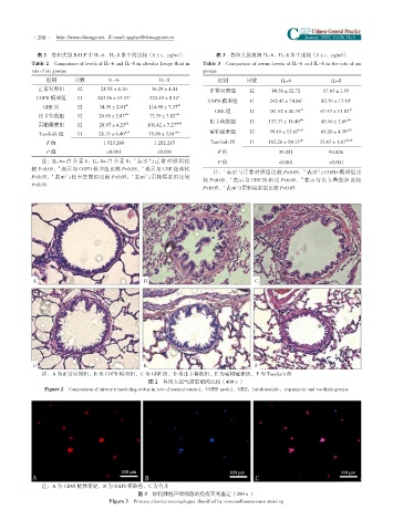


           A                                 B                                 C


           D                                 E                                 F
              注:A 为正常对照组,B 为 COPD 模型组,C 为 GBE 组,D 为比卡鲁胺组,E 为雷帕霉素组,F 为 Taselisib 组
                                              图 2 各组大鼠气道重塑的比较(400×)
               Figure 2 Comparison of airway remodeling status in rats of normal control,COPD model,GBE,bicalutamide,rapamycin and taselisib groups


              注:A 为 CD68 抗体鉴定,B 为 DAPI 核染色,C 为合并
                                           图 3 原代肺泡巨噬细胞的免疫荧光鉴定(200×)
                                  Figure 3 Primary alveolar macrophages identified by immunofluorescence staining
   44   45   46   47   48   49   50   51   52   53   54