中国全科医学 ›› 2024, Vol. 27 ›› Issue (24): 2976-2981.DOI: 10.12114/j.issn.1007-9572.2023.0679

• 论著 • 上一篇    下一篇

泛免疫炎症值、全身免疫炎症指数与急性冠脉综合征患者易损斑块的相关性研究

和军辉, 万大国*(), 董静, 张娟   

  1. 450000 河南省郑州市,郑州大学第二附属医院心血管内科
  • 收稿日期:2023-07-27 修回日期:2024-01-06 出版日期:2024-08-20 发布日期:2024-05-08
  • 通讯作者: 万大国

  • 作者贡献:

    和军辉进行研究设计、数据分析、撰写论文;董静、张娟负责文章审校;万大国负责最终版本修订,对论文整体负责。

  • 基金资助:
    河南省医学科技攻关计划(联合共建)项目(LHGJ20220438)

Correlation Analysis between Pan-immune Inflammatory Value, Systemic Immune-inflammatory Index, and Vulnerable Plaques in Patients with Acute Coronary Syndrome

HE Junhui, WAN Daguo*(), DONG Jing, ZHANG Juan   

  1. Department of Cardiology, the Second Affiliated Hospital of Zhengzhou University, Zhengzhou 450000, China
  • Received:2023-07-27 Revised:2024-01-06 Published:2024-08-20 Online:2024-05-08
  • Contact: WAN Daguo

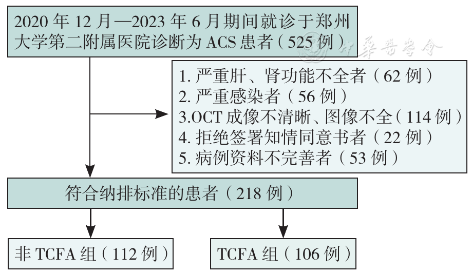
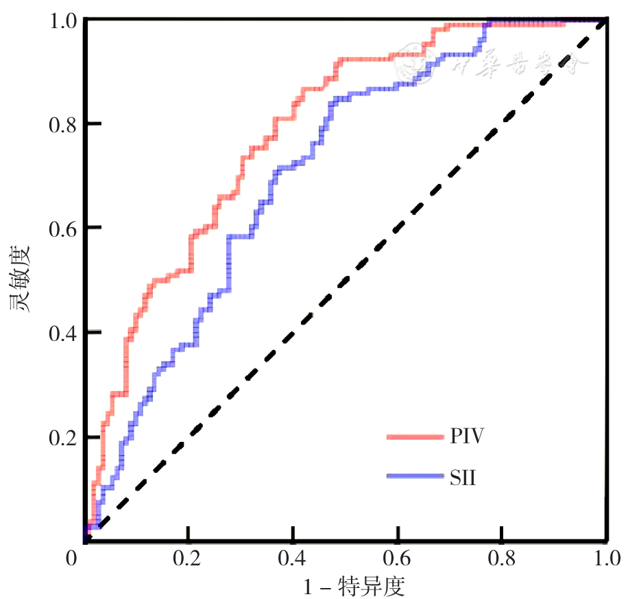
摘要: 背景 泛免疫炎症值(PIV)、全身免疫炎症指数(SII)被认为是评估动脉粥样硬化性心血管疾病风险的新型炎症指标,然而较少有研究探讨PIV、SII对动脉粥样硬化斑块的影响。 目的 通过光学相干断层成像技术(OCT)探讨急性冠脉综合征(ACS)患者PIV、SII与冠状动脉粥样硬化易损斑块的关系。 方法 回顾性纳入2020年12月—2023年6月就诊于郑州大学第二附属医院心内科的ACS患者525例为研究对象,患者均接受冠状动脉造影及OCT检查,根据薄纤维帽粥样硬化斑块(TFCA)诊断标准分为非TCFA组(112例)和TCFA组(106例)。收集资料并进行分析。采用多因素有序Logistic回归分析探究TCFA的影响因素。绘制PIV、SII诊断TCFA的受试者工作特征曲线(ROC曲线),计算ROC曲线下面积(AUC)。基于PIV的最佳截断值将患者分为低PIV组(PIV<337.86,79例)和高PIV组(PIV≥337.86,139例),同时基于SII的最佳截断值将患者分为低SII组(SII<775.63,74例)和高SII组(SII≥775.63,144例)。 结果 TCFA组高血压、糖尿病比例、泛PIV、SII、C反应蛋白(CRP)、有吸烟史比例、术前收缩压高于非TCFA组(P<0.05)。多因素Logistic回归分析结果显示PIV(OR=1.015,95%CI=1.010~1.020,P<0.001)、SII(OR=1.005,95%CI=1.003~1.007,P<0.001)为发生TCFA的危险因素。ROC曲线结果显示PIV、SII诊断TCFA的AUC分别为0.785(95%CI=0.725~0.845,P<0.001)、0.707(95%CI=0.639~0.776,P<0.001)。高PIV组巨噬细胞浸润、点状钙化、易损斑块比例高于低PIV组(P<0.05);高SII组巨噬细胞浸润、微通道、易损斑块比例高于低SII组(P<0.05)。高PIV组纤维帽厚度低于低PIV组,最大脂质斑块角、平均脂质斑块角度、脂质斑块长度、脂质指数高于低PIV组(P<0.05),高SII组纤维帽厚度低于低SII组,最大脂质斑块角、平均脂质斑块角度、脂质斑块长度、脂质指数高于低SII组(P<0.05)。 结论 高水平PIV、SII可能与ACS患者易损斑块的发生率相关。PIV、SII水平在评估ACS患者冠状动脉粥样硬化易损斑块特征及易损性方面具有一定价值。

关键词: 冠状动脉疾病, 冠状动脉粥样硬化, 急性冠脉综合征, 光学相干断层成像技术, 泛免疫炎症值, 全身免疫炎症指数

Abstract:

Background

The pan-immune inflammatory value (PIV) and systemic immune-inflammatory index (SII) are considered novel inflammatory markers for assessing the risk of atherosclerotic cardiovascular diseases. However, few studies have confirmed the impact of PIV and SII on atherosclerotic plaques.

Objective

To explore the relationship between PIV, SII, and vulnerable atherosclerotic plaques in patients with acute coronary syndrome (ACS) using optical coherence tomography (OCT) .

Methods

This retrospective study included 525 ACS patients treated at the Second Affiliated Hospital of Zhengzhou University from December 2020 to June 2023. All patients underwent coronary angiography and OCT imaging. Patients were further categorized into low PIV (<337.86, 79 cases) and high PIV (≥337.86, 139 cases) groups based on the optimal PIV cutoff value, as well as into low SII (<775.63, 74 cases) and high SII (≥775.63, 144 cases) groups based on the optimal SII cutoff value. Data were collected and analyzed. Multifactorial ordinal Logistic regression analysis was used to explore the influencing factors of TCFA. Receiver operating characteristic (ROC) curves were plotted for PIV and SII in diagnosing TCFA, and the area under the curve (AUC) was calculated. Based on the diagnostic criteria for thin-cap fibroatheroma (TCFA), patients were divided into a non-TCFA group (112 cases) and a TCFA group (106 cases) .

Results

The TCFA group had higher proportions of hypertension, diabetes, pan-PIV, SII, C-reactive protein (CRP), smoking history, and preoperative systolic pressure compared to the non-TCFA group (P<0.05). Multifactorial Logistic regression analysis showed that PIV (OR=1.015, 95%CI=1.010-1.020, P<0.001) and SII (OR=1.005, 95%CI=1.003-1.007, P<0.001) were risk factors for TCFA. ROC curve results indicated that the AUCs for PIV and SII in diagnosing TCFA were 0.785 (95%CI=0.725-0.845, P<0.001) and 0.707 (95%CI=0.639-0.776, P<0.001), respectively. The high PIV group showed higher rates of macrophage infiltration, punctate calcification, and vulnerable plaques than the low PIV group (P<0.05). The high SII group showed higher rates of macrophage infiltration, microchannels, and vulnerable plaques than the low SII group (P<0.05). The high PIV group had thinner fibrous caps, larger maximum lipid core angle, average lipid core angle, lipid core length, and lipid index compared to the low PIV group (P<0.05), and the high SII group had thinner fibrous caps, larger maximum lipid core angle, average lipid core angle, lipid core length, and lipid index compared to the low SII group (P<0.05) .

Conclusion

High levels of PIV and SII may be associated with the incidence of vulnerable plaques in patients with ACS. PIV and SII levels have potential value in assessing the characteristics and vulnerability of coronary atherosclerotic plaques in ACS patients.

Key words: Coronary artery disease, Coronary atheroscleroses, Acute coronary syndrome, Optical coherence tomography, Pan-immune-inflammation value, Systemic immune-inflammation index